The Role of Medical Imaging in Modern Treatment Procedures
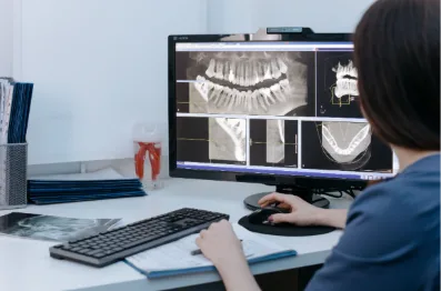
Medical imaging has changed the way healthcare professionals diagnose diseases, plan treatments, and perform medical procedures. Technologies such as ultrasound, computed tomography (CT), magnetic resonance imaging (MRI), and fluoroscopy allow physicians to examine internal structures without making large surgical incisions.
These technologies are no longer limited to identifying medical conditions. They also help physicians guide instruments, deliver treatments, and monitor results with greater precision.
For patients, this development has created more opportunities to receive targeted treatments that may involve less physical disruption than conventional surgery.
From treating blocked blood vessels to managing certain tumors and chronic medical conditions, medical imaging has become an important part of modern healthcare.
Understanding how imaging technology supports treatment can help patients make more informed decisions when discussing their medical options.
Understanding Medical Imaging and Its Importance
Medical imaging refers to technologies that produce visual representations of structures inside the human body. Healthcare professionals use these images to examine organs, bones, muscles, blood vessels, and surrounding tissues.
Different imaging techniques provide different types of information.
For example, X-rays are commonly used to examine bones and certain chest conditions, while ultrasound can visualize soft tissues and blood flow.
CT scans create detailed cross-sectional images, making them useful for examining complex internal structures.
MRI uses magnetic fields and radio waves to produce detailed images of organs and soft tissues without using ionizing radiation.
The selection of an imaging technique depends on the medical condition, the area being examined, and the information required.
Accurate imaging helps physicians understand the location and extent of disease before recommending treatment.
How Medical Imaging Supports Accurate Diagnosis
One of the most important functions of medical imaging is identifying conditions that physical examinations alone cannot fully evaluate.
Symptoms such as abdominal pain, swelling, unexplained bleeding, or persistent joint discomfort may have several possible causes.
Imaging examinations can help healthcare professionals distinguish between these conditions and determine whether additional testing is necessary.
For example, ultrasound may reveal uterine fibroids, while MRI can provide detailed information about their location and size.
Similarly, vascular imaging can help identify narrowed blood vessels, abnormal circulation, or other conditions affecting blood flow.
Early identification of abnormalities may allow physicians to recommend appropriate treatment before complications develop.
However, imaging results must be interpreted alongside medical history, symptoms, laboratory findings, and physical examination.
An abnormal image does not always mean treatment is necessary, and normal imaging results do not automatically rule out every medical condition.
The Growing Importance of Image-Guided Treatments
One of the most significant developments in modern medicine is the use of imaging technology during treatment procedures.
Instead of relying entirely on direct surgical visibility, physicians can use live images to guide small instruments toward specific areas inside the body.
This approach is especially important in interventional radiology, a medical specialty that uses imaging guidance to perform minimally invasive diagnostic and therapeutic procedures.
Specialists may use ultrasound, CT, or fluoroscopy to position needles, catheters, and other medical devices.
For example, facilities such as Image Guided Therapy specialize in minimally invasive procedures that use advanced medical imaging to guide targeted treatments. These approaches include embolization procedures for selected vascular and nonvascular medical conditions.
Image guidance allows physicians to identify relevant anatomical structures and monitor the position of instruments throughout treatment.
Depending on the procedure, this may reduce the need for large incisions and allow patients to recover more quickly than they would after traditional open surgery.
However, not every condition is suitable for an image-guided procedure, and treatment recommendations must consider each patient’s medical circumstances.
Common Medical Imaging Technologies Used During Treatment
Several imaging technologies play an important role in guiding modern medical procedures.
Ultrasound Guidance
Ultrasound uses high-frequency sound waves to produce images of internal tissues.
Because it provides real-time images without ionizing radiation, it is widely used during biopsies, fluid drainage, vascular access, and other procedures.
Physicians can observe the movement of instruments while avoiding nearby blood vessels and sensitive structures.
CT-Guided Procedures
CT imaging provides detailed cross-sectional views of the body.
It is particularly useful when physicians need to locate abnormalities deep within tissues or organs.
CT guidance may be used during certain biopsies, drainage procedures, and tumor treatments.
Fluoroscopy
Fluoroscopy uses continuous or pulsed X-rays to create moving images of internal structures.
It is especially valuable during procedures involving blood vessels.
Physicians can observe the movement of catheters and contrast material as they navigate through the vascular system.
MRI Guidance
MRI provides detailed soft-tissue images and does not use ionizing radiation.
Although MRI-guided procedures require specialized equipment and are not appropriate for every intervention, they can support selected treatments where detailed tissue visualization is important.
Each technology has advantages and limitations, making appropriate selection essential.
The Role of Imaging in Minimally Invasive Procedures
Minimally invasive procedures often use small openings in the skin instead of large surgical incisions.
Medical imaging helps physicians navigate instruments through these openings while identifying the intended treatment area.
For example, a physician performing an embolization procedure may insert a catheter into a blood vessel and guide it toward a specific artery.
Once the catheter reaches the appropriate location, the physician can deliver materials designed to reduce or block blood flow through the targeted vessel.
Imaging helps confirm catheter placement and monitor treatment delivery.
This approach can be useful for selected conditions involving abnormal blood vessels, tumors, or other vascular problems.
Compared with traditional open surgery, some minimally invasive procedures may involve shorter hospital stays, reduced postoperative discomfort, and faster recovery.
However, outcomes depend on the patient’s condition, the procedure performed, and the expertise of the treating team.
How Imaging Helps Treat Uterine Fibroids
Uterine fibroids are noncancerous growths that develop within or around the uterus.
While many fibroids cause no symptoms, others can produce heavy menstrual bleeding, pelvic pressure, and discomfort.
Medical imaging plays an important role in identifying fibroids and evaluating their size and location.
Ultrasound is commonly used during the initial assessment, while MRI may provide additional information when needed.
For selected patients, uterine fibroid embolization offers a minimally invasive treatment option.
During this procedure, an interventional radiologist uses imaging guidance to position a catheter in arteries supplying the fibroids.
Small particles are then delivered to reduce blood flow to the growths.
Over time, the fibroids may shrink, helping relieve certain symptoms.
However, treatment selection depends on fibroid characteristics, reproductive plans, and individual medical needs.
Image-Guided Procedures for Vascular Conditions
Blood vessels supply oxygen and nutrients throughout the body. When circulation becomes obstructed or abnormal, patients may develop pain, swelling, or other complications.
Medical imaging helps physicians examine the vascular system and identify areas requiring treatment.
Doppler ultrasound can assess blood flow, while CT angiography and other vascular imaging techniques provide detailed information about blood vessel anatomy.
During some procedures, physicians use fluoroscopy to guide catheters through arteries or veins.
Image-guided interventions may include angioplasty, stent placement, embolization, and treatments designed to remove or dissolve certain blood clots.
These procedures do not replace surgery in every situation.
Their suitability depends on the location of the vascular problem, its severity, and the patient’s overall health.
Medical Imaging in Cancer Treatment
Medical imaging also contributes to cancer care by helping physicians locate tumors, evaluate disease progression, and plan treatment.
CT, MRI, ultrasound, and other imaging technologies provide information about tumor size, location, and relationships with surrounding structures.
Certain cancer treatments use imaging to guide instruments directly toward tumors or the blood vessels supplying them.
For example, image-guided ablation techniques may destroy selected tumors using heat, extreme cold, or other forms of energy.
In some liver cancer treatments, physicians use catheter-based procedures to deliver therapeutic materials to targeted blood vessels.
These approaches may be appropriate for certain patients depending on cancer type, stage, and treatment goals.
They are typically considered as part of a broader treatment plan developed by a multidisciplinary medical team.
Benefits and Limitations of Image-Guided Treatments
Medical imaging can improve procedural planning and allow physicians to target specific structures with greater accuracy.
Potential advantages of image-guided treatment include:
- Smaller incisions for certain procedures
- More precise placement of instruments
- Reduced disruption to surrounding tissues
- Shorter recovery periods in selected cases
- Opportunities for outpatient treatment
- Real-time monitoring during procedures
Despite these benefits, image-guided treatments still carry risks.
Potential complications vary by procedure and may include bleeding, infection, blood vessel injury, allergic reactions to contrast agents, or unintended damage to surrounding tissues.
Some imaging methods also expose patients to ionizing radiation.
The Society of Interventional Radiology explains how imaging technologies support minimally invasive procedures and targeted medical treatments.
Patients should discuss the expected benefits, possible complications, alternative treatments, and recovery requirements before undergoing an intervention.
How Medical Imaging Supports Treatment Monitoring
Medical imaging remains valuable after treatment is completed.
Follow-up examinations can help physicians determine whether a procedure achieved its intended purpose and whether additional care is required.
For example, imaging may be used to assess changes in tumor size, evaluate blood flow after vascular treatment, or monitor the response of uterine fibroids following embolization.
The type and frequency of follow-up imaging depend on the medical condition and treatment performed.
Regular monitoring can help identify complications or incomplete treatment responses.
However, follow-up care should focus on both imaging findings and the patient’s symptoms, rather than relying exclusively on scan results.
The Future of Medical Imaging in Healthcare
Medical imaging continues to evolve as healthcare professionals develop more sophisticated ways to visualize and treat disease.
Artificial intelligence is being investigated for applications such as image interpretation, identifying abnormalities, and supporting procedural planning.
Three-dimensional imaging and advanced navigation systems may also help physicians understand complex anatomy and plan certain interventions more effectively.
These developments have the potential to improve efficiency and precision, although new technologies require clinical validation before widespread adoption.
The future of medical imaging will likely involve closer integration between diagnostic information, treatment planning, and minimally invasive procedures.
Conclusion
Medical imaging has become an essential part of modern healthcare, supporting everything from early diagnosis to complex treatment procedures.
Technologies such as ultrasound, CT, MRI, and fluoroscopy help physicians understand internal anatomy, identify medical conditions, and guide instruments during targeted interventions.
The growing use of image-guided procedures has expanded treatment options for patients with certain vascular conditions, uterine fibroids, tumors, and other medical problems.
Although these techniques offer important advantages, they are not suitable for every patient or condition.
Understanding the benefits, risks, and limitations of medical imaging allows patients to participate more actively in treatment decisions and work with healthcare professionals to select appropriate care.