
How a Fundus Camera for Optometry Practice Enhances Early Detection of Retinal Diseases
First, a flash of light—and then panic.
A patient blinks, stunned. “What was that?”
You, the optometrist, smile. “Just taking a look at your retina.”
What you don’t say? That flash might’ve just caught the earliest signs of diabetic retinopathy. Or glaucoma. Or age-related macular degeneration—long before it dares show itself.
That, friends, is the magic of the fundus camera for optometry practice. No tarot cards. No guesswork. Just retinal intel delivered with the speed of a shutter click.
Silent diseases don’t play fair.
Here’s the brutal truth: most retinal conditions don’t knock first. They sneak in. Take a seat. Set up camp.
By the time the patient says, “my vision seems off,” it’s often too late to undo the damage.
- Diabetic retinopathy? Already working its way through the vasculature.
- Glaucoma? Nibbling at the optic nerve like it owns the place.
- AMD? You might catch it if you’re lucky. Or if you’re using a fundus camera.
So why are some optometrists still relying on standard ophthalmoscopy alone? It’s like bringing a fork to a fencing match.
Early detection isn’t flashy—it’s life-changing.
Yes, it’s cool tech. High-res imaging. Color, depth, vascular structure.
But that’s not the story.
The story is the 44-year-old with type 2 diabetes who gets retinal imaging during her routine exam—no symptoms, no complaints. And there it is: a cluster of microaneurysms forming like storm clouds. You catch it. You refer. Her vision is saved.
That’s the win.
That’s the ROI.
And that’s just Tuesday with a fundus camera.
Fact: Nearly 1 in 3 diabetics has retinopathy, often without symptoms.
Source: CDC (Yes, we’re citing government studies now. It’s that serious.)
Fast. Painless. Staff-friendly. Patient-impressing.
Let’s talk workflow—because no one wants another gadget that slows down the day.
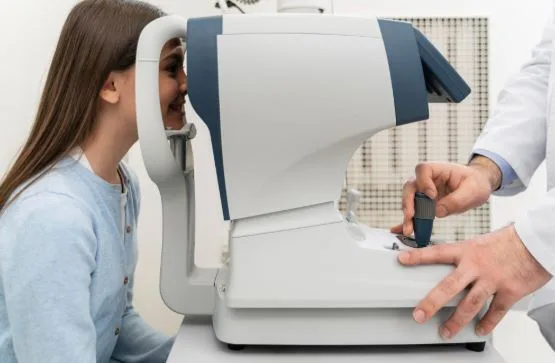
Non-mydriatic fundus cameras are fast. Pop, click, done.
- No dilation needed? Check.
- Minimal training? Check.
- Tech does the image, doc reads it later? Beautiful.
Even better? Patients love seeing their retinas. You’d be surprised how motivational a zoomed-in macula can be. It’s no Mona Lisa, but it gets people thinking about sugar intake real quick.
Documentation that doesn’t get dusty.
Baseline images? Stored.
Last year’s comparison? One click away.
Sharing with a retinal specialist or PCP? Digital file, sent in seconds.
And for the overachievers: some systems even offer AI-driven anomaly detection. (Yes, your camera is smarter than Carl, the guy who forgets to refill the contact lens solution.)
Let’s get blunt—this tech pays for itself.
Fundus imaging isn’t just good medicine. It’s good business.
You’re not upselling glasses. You’re documenting pathology. Billing appropriately. Providing actual preventive care that insurance can recognize.
It’s the kind of upgrade that builds patient trust, not just revenue.
According to The American Journal of Ophthalmology, using annual fundus imaging increased detection of treatable retinal conditions by 62%.
That’s not a statistic. That’s a wake-up call.
So why isn’t every practice using one?
Let’s not pretend budget isn’t a concern. It is.
But when you’re weighing a piece of equipment that literally prevents blindness, you might want to reframe the cost as “not-being-sued-for-missing-a-glaucoma-diagnosis money.”
Kidding. (Mostly.)
TL;DR: Fundus cameras are the future of standard care.
You can’t see what you don’t look for.
You can’t catch what you don’t image.
You can’t protect what you don’t prioritize.
Modern optometry isn’t just about refractions and retinal blur. It’s about being proactive, predictive, and yes—technically prepared.