Keratoconus: What It Is, Why It Happens, and What You Can Do About It
Most people have never heard of keratoconus until the day their eye doctor says those four syllables and everything changes. Suddenly, blurry vision that glasses couldn’t fix makes sense. The halos around streetlights at night make sense. The frustration of constantly updating your prescription, only to still not see clearly, makes sense.
Keratoconus is more common than many people realize, affecting roughly 1 in every 2,000 people worldwide. And yet it remains widely misunderstood, often mistaken for ordinary nearsightedness or astigmatism for years before a correct diagnosis is made. The good news is that with the right knowledge and the right care team, keratoconus is very manageable. People with the condition go on to live full, active lives with excellent vision.
This article walks you through everything you need to know: what keratoconus actually is, what causes it, how to recognize its symptoms, and the full range of treatment options available today.
What Is Keratoconus?
To understand keratoconus, you first need a basic picture of how the eye works.
The cornea is the clear, dome-shaped window at the very front of your eye. Its job is to focus incoming light onto the retina at the back of the eye, which then sends signals to your brain to form an image. For this process to work properly, the cornea needs to maintain a smooth, rounded shape.
In keratoconus, the structural integrity of the cornea gradually breaks down. The collagen fibers that normally hold the cornea in its rounded shape weaken over time, causing the cornea to thin and begin to bulge outward, taking on a cone-like shape. This is where the name comes from: kerato (cornea) + conus (cone).
As the cornea becomes increasingly irregular in shape, it can no longer focus light accurately. The result is distorted, blurry vision that standard eyeglass prescriptions struggle to correct, because glasses are designed for regularly shaped corneas, not cone-shaped ones.
Keratoconus typically begins in the teenage years or early twenties and can progress for a decade or more before stabilizing. In most cases it affects both eyes, though often unevenly. One eye may be more advanced than the other.
What Causes Keratoconus?
Researchers have been studying keratoconus for decades, and while the full picture is still emerging, several factors are known to contribute to its development.
Genetics
Keratoconus runs in families. If a parent or sibling has the condition, your own risk is meaningfully higher than the general population. Studies suggest that between 10 and 20 percent of keratoconus patients have a family member with the condition. If you have a close relative with keratoconus, it’s worth discussing regular corneal screening with your eye doctor, even if your vision currently seems fine.
Eye Rubbing
This is one of the most well-established environmental risk factors for keratoconus, and also one of the most controllable. Chronic, vigorous eye rubbing, the kind associated with allergies, habit, or even aggressive eye makeup removal, creates repeated mechanical stress on the cornea. Over time, this stress is believed to accelerate the breakdown of collagen fibers.
People who rub their eyes frequently, particularly those with atopic disease (eczema, hay fever, or allergic conjunctivitis), are at significantly higher risk. If you have keratoconus or a family history of it, stopping eye rubbing is one of the most important lifestyle changes you can make.
Connective Tissue Disorders
Keratoconus is associated with several systemic conditions that affect connective tissue, including Down syndrome, Ehlers-Danlos syndrome, and Marfan syndrome. The connection makes intuitive sense. If the body’s connective tissue is generally more fragile, this vulnerability may extend to the collagen that supports the cornea.
Oxidative Stress
Some research points to an imbalance of antioxidants in keratoconic corneas, making them more vulnerable to damage from free radicals. This is still an active area of investigation and may help explain why the corneal tissue degrades over time even in the absence of obvious mechanical trauma.
Contact Lens Wear (Poorly Fitted)
Wearing poorly fitted rigid contact lenses over long periods has historically been associated with corneal warpage, though researchers continue to debate the precise relationship between contact lens wear and keratoconus progression. What is clear is that improperly fitted lenses on an already compromised cornea can cause additional stress. This is why working with a specialty contact lens clinic in Kansas that has the right experience and equipment matters so much from the very beginning.
Recognizing the Symptoms
Keratoconus is a progressive condition, which means symptoms tend to worsen gradually. In its early stages it can be very easy to dismiss or misattribute. Here are the warning signs to watch for.
Frequent Prescription Changes
One of the earliest and most telling signs of keratoconus is a prescription that keeps shifting, never staying stable for more than a year or two. If you or your child is updating glasses or contact lens prescriptions frequently without ever achieving fully clear vision, this warrants a closer look at the cornea.
Blurred or Distorted Vision
As the cornea changes shape, vision becomes increasingly distorted. Straight lines may appear wavy or bent. Text can look smeared or ghosted. Standard corrective lenses may improve vision somewhat but rarely bring it to a sharp, clear level.
Sensitivity to Light and Glare
Many people with keratoconus describe significant light sensitivity, particularly in bright sunlight. Night driving can become especially difficult as headlights and streetlights appear to scatter into starburst patterns or halos.
Multiple Ghost Images (Monocular Diplopia)
Even when covering one eye, a person with keratoconus may see multiple overlapping images of a single object. This is a distinctive sign of irregular astigmatism, the kind produced by a misshapen cornea, and it is quite different from the double vision caused by eye alignment problems.
Eye Strain and Headaches
The brain works overtime trying to make sense of the distorted signals coming from a keratoconic eye. This extra effort can manifest as eye strain, fatigue, or headaches, particularly after reading or screen use.
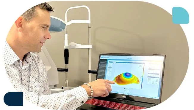
If several of these symptoms sound familiar, the next step is a comprehensive eye exam that includes corneal topography, which is a detailed mapping of the cornea’s surface shape. This test is not always included in a routine eye exam, so it’s worth specifically requesting if you have concerns.
How Keratoconus Is Diagnosed
A standard eye chart test or refraction cannot diagnose keratoconus. What’s needed is a more detailed look at the cornea itself.
Corneal topography creates a color-coded map of the corneal surface, revealing irregular patterns that are characteristic of keratoconus. A healthy cornea produces a fairly uniform, round map. A keratoconic cornea shows a skewed, asymmetric pattern often described as a “football” or “sagging” shape.
Corneal tomography (such as a Pentacam scan) goes a step further, producing a three-dimensional image that measures both the front and back surfaces of the cornea, as well as its thickness at various points. This allows detection of very early keratoconus, sometimes before symptoms even appear.
Pachymetry measures corneal thickness. A thinning cornea, particularly when combined with topographic irregularity, strengthens a keratoconus diagnosis.
Early detection matters because treatment options and outcomes are significantly better when the condition is caught before it has progressed far.
Treatment Options: From Early to Advanced
The treatment approach for keratoconus depends on how far the condition has progressed. Here is a full overview of the options currently available.
1. Glasses and Soft Contact Lenses (Early Stage)
In the mildest cases of keratoconus, ordinary glasses or soft contact lenses may provide adequate vision. However, as the cornea becomes more irregular, these options quickly become insufficient. Most patients eventually need something more specialized.
2. Corneal Cross-Linking (CXL)
Corneal cross-linking is the only treatment currently proven to halt the progression of keratoconus. It does not reverse existing damage, but it can stop things from getting worse.
The procedure involves applying riboflavin (vitamin B2) eye drops to the cornea and then activating them with ultraviolet light. This creates new bonds between the collagen fibers in the cornea, strengthening its structure. Cross-linking is typically recommended when progression is documented, meaning the cornea is measurably changing over time, particularly in younger patients whose keratoconus is more likely to continue advancing.
Recovery takes several weeks, and mild discomfort and blurred vision are expected in the short term. The long-term results are generally very good. Most patients see their progression stop, and some experience modest improvement in corneal shape over time.
3. Gas Permeable (GP) Contact Lenses
Also called rigid gas permeable or RGP lenses, these hard lenses create a smooth, regular optical surface over the irregular cornea, dramatically improving vision. Because they don’t conform to the cornea’s distorted shape the way soft lenses do, they can provide much sharper correction.
The tradeoff is comfort. Rigid lenses take longer to adapt to, and fitting them on a keratoconic cornea requires a skilled and experienced practitioner. When fitted well, however, many patients wear them comfortably for years.
4. Scleral Lenses
Scleral lenses are large-diameter rigid lenses that vault completely over the cornea and rest on the white of the eye (the sclera) instead. Because they never touch the cornea itself, they avoid the discomfort issues that can arise with smaller rigid lenses on sensitive or irregular corneas.
The space between the lens and the cornea is filled with preservative-free saline, creating a smooth liquid layer that acts as a perfect optical surface. This means that even highly irregular corneas, including advanced keratoconus, post-surgical eyes, and corneas with scarring, can achieve excellent vision with scleral lenses.
Scleral lens fitting for keratoconus requires specialized equipment and a practitioner who works with these lenses day in and day out. When done well, the results can be genuinely life-changing. Many patients who spent years struggling with blurry, distorted vision finally see clearly for the first time after being fitted properly with sclerals.
5. Hybrid Lenses
Hybrid lenses combine a rigid center with a soft outer skirt. The rigid center provides the optical clarity of a GP lens, while the soft skirt improves comfort and centration. They are a good middle-ground option for patients who find fully rigid lenses uncomfortable but aren’t achieving adequate vision with soft lenses.
6. EyePrintPRO and Impression-Based Lenses
For the most complex or irregular corneas, including eyes with extreme keratoconus, significant scarring, or previous surgeries, impression-based lenses represent the cutting edge of custom fitting. A mold or detailed scan of the eye’s actual surface is taken, and a lens is custom-fabricated to match it precisely. This level of customization can achieve comfortable, clear vision even in cases where all other options have been exhausted.
7. Corneal Transplant (Penetrating Keratoplasty or DALK)
When the cornea has thinned to the point where contact lenses can no longer provide adequate vision, or if significant scarring has developed, a corneal transplant may be considered. In this procedure, the damaged corneal tissue is replaced with donor tissue.
Penetrating keratoplasty (PK) replaces the full thickness of the cornea, while Deep Anterior Lamellar Keratoplasty (DALK) replaces only the outer layers, preserving the patient’s own inner lining. DALK carries a lower risk of rejection and is generally preferred when it is technically feasible.
It’s important to note that even after a successful corneal transplant, most patients still need specialty contact lenses to achieve their best vision. The transplanted cornea rarely produces a perfectly regular surface, which is why access to experienced keratoconus contact lens specialists remains important even post-surgery.
Living With Keratoconus
A keratoconus diagnosis can feel overwhelming at first, but the condition is highly manageable with the right care. Here are some practical takeaways for living well with it.
Stop rubbing your eyes. This is non-negotiable. Address underlying allergies with antihistamine drops, use cold compresses for itch relief, and break the habit however you can.
Get regular monitoring. Even stable keratoconus should be checked with corneal topography at least once a year, so any changes are caught early.
Work with a specialist. General optometrists are excellent for routine care, but keratoconus management, especially contact lens fitting, is best handled by practitioners with specific expertise and the right technology. The difference in outcomes can be dramatic.
Don’t give up on good vision. Even advanced keratoconus can often be corrected to 20/20 or close to it with the right lens design. It may take patience and multiple fitting sessions, but the results are life-changing for many patients.
Final Thoughts
Keratoconus is a condition that asks more of patients than ordinary vision problems. It requires more vigilance, more specialized care, and often more patience. But it is also a condition with a remarkable array of effective solutions. From cross-linking to scleral lenses to impression-based custom designs, the field of keratoconus management has advanced enormously in recent years.
If you or someone you love is struggling with vision that glasses can’t fix, or has received a keratoconus diagnosis and isn’t sure what comes next, the most important step is connecting with a specialist who truly understands the condition. The Contact Lens Institute of Kansas focuses exclusively on complex contact lens cases, including keratoconus, and has helped patients achieve clear, comfortable vision when other options had failed. The right practitioner, the right technology, and the right lens can make all the difference between struggling to see and experiencing the world with real clarity again.