
Streamlining Sample-to-Image Workflows With Smart Tools
A brilliant image in a research paper tells a story. It captures months of work in a single frame. But the journey to get that picture is rarely straightforward. It is often a messy, multi-step obstacle course. Samples move from hands to tubes. They go from plates to slides. Each transfer is a chance for error. Each delay risks degradation.
This “sample-to-image” gauntlet is where great science often stumbles. Thankfully, a new generation of smart tools is changing the game. They are creating a seamless, reliable highway from preparation to publication.
Unlocking Your Biggest Investments
Labs invest heavily in imaging power. They acquire incredible instruments to see the unseen. But an instrument is only as good as the sample placed under its lens. A poorly prepared specimen wastes that potential. It wastes something else, too: time and money.
Smart workflow tools ensure your sample is perfect every time. This is how you truly maximize the return on a major capital outlay, like your confocal microscope price. The goal is not just to take images. It is to extract perfect, publishable data on the first try. Streamlining the steps before imaging makes that possible.
The Labeling Revolution
Let’s start at the very beginning. Sample tracking is a nightmare. Handwritten labels smudge. Tubes get mixed up. This creates a cascade of confusion. Smart tools solve this. Digital sample management platforms use barcodes. Everything gets scanned. Every tube, every plate has a digital twin.
This creates an unbreakable chain of custody. You always know what a sample is. You know its entire history. This eliminates tragic mix-ups. It turns sample management from a chore into a silent, automated background process.
Preparation Without the Guesswork
Sample prep itself is full of variables. Protocol steps are complicated. Timings are precise. Human memory is imperfect. Smart protocol assistants guide technicians through each step. They use clear on-screen instructions.
Some systems even integrate with automated liquid handlers. The software tells the robot exactly what to do next. This removes human deviation from the equation. It ensures every sample is treated with identical care. This consistency is the bedrock of reproducible, high-quality staining and processing.
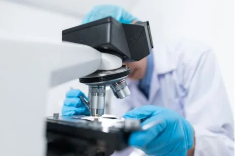
The Intelligent Imaging Session
Now, the sample is ready. The old way involved manual navigation. A scientist hunts for the right cells. This takes forever. It is also subjective. Smart microscopy software changes this. It uses advanced algorithms. It can automatically find regions of interest. It can focus perfectly. It can even adjust exposure settings on the fly.
Some systems perform entire imaging routines unattended. They work through the night. This saves hours of human labor. It also captures data a tired person might miss at midnight.
From Pixels to Answers, Faster
The image is captured. The work is not done. Analysis is the next bottleneck. Manually outlining cells is tedious. Counting particles by eye is painful. Smart analysis tools use artificial intelligence. They learn what you are looking for. They can segment cells automatically. They can quantify fluorescence intensity across thousands of images in minutes.
This turns a raw picture into quantitative data almost instantly. The insight is no longer buried in pixels. It is presented in clear graphs and statistics, ready for interpretation.
Connecting the Digital Dots
The real magic happens when these tools talk to each. A truly streamlined workflow is fully integrated. The sample management system knows a protocol is complete. It alerts the microscope software. The microscope acquires the images. It then sends them directly to the analysis module.
This creates a closed-loop, digital pipeline. Data never sits in a forgotten folder. It flows automatically to the next stage. This integration eliminates manual file handling. It prevents version errors. It makes the entire process traceable and audit-ready.
The Tangible Payoff
The benefits are immediate and real. Labs experience a dramatic drop in failed experiments. They see a massive increase in throughput. Researchers get their data faster. They have more confidence in its quality.
Most importantly, scientists are freed from repetitive tasks. They can focus on experimental design and deep data interpretation. Their expertise is applied to thinking, not pipetting or clicking.
A Smarter Way to Work
Adopting these tools is an investment in smoothness. It is a commitment to removing friction from science. The path from a raw sample to a powerful image becomes predictable. It becomes reliable. This streamlined approach does more than save time. It elevates the quality and impact of the research itself.
In a competitive world, the smartest tool isn’t always the microscope. Sometimes, it’s the invisible system that ensures the microscope always has something perfect to see.