What is a 3D microscope?
A standard microscope can only show you a flat, two-dimensional image, leaving out crucial details like depth, height, and surface contours. Enter the 3D microscope—a powerful tool that lets you explore specimens in full three-dimensional detail, offering a more complete view than what a 2D microscope can provide.
In this article, you’ll discover exactly what a 3D microscope is, how it works, and the different types available, including stereo, digital, confocal, and holographic systems. You’ll also learn about the key benefits of using 3D microscopy, such as improved precision in measuring height and depth, and how these tools can be applied across fields from biology to materials science. Keep reading to find out how 3D microscopes can take your analysis to the next level.
| Microscope Type | Primary “3D” Output | Best For | Typical Use Cases | Cost Range |
| Stereo Microscope | True, Live 3D Depth Perception (via eyepieces) | Manual Inspection & Manipulation (low power) | Soldering, Dissection, Jewelry, Watch Repair | Entry-level to Mid |
| 3D Digital Microscope | Computer-Generated 3D Models (software-based) | Surface Analysis, Measurement, Documentation | Quality Control, Failure Analysis, Forensics | Mid to High |
| Confocal Microscope | Optical Sectioning for Internal 3D Structure | Advanced Scientific Research (high resolution) | Cell Biology, Live Imaging, Material Science | High to Very High |
| 3D Electron Microscope | Ultra-High Resolution 3D Reconstructions | Nanoscale Structure Analysis (research) | Materials Science, Nanotechnology, Life Sciences | Very High (Research) |
Understanding the Essence of 3D Microscopy
At its core, a 3D microscope overcomes the limitations of a traditional 2D microscope by capturing information about a specimen’s height and depth. This allows you to visualize its true shape, inspect surface features, or even explore internal structures that would remain hidden in a flat image.
The Power of Perceiving Depth
Imagine trying to understand the topography of a mountain range from a flat map. It gives you some information, but it pales in comparison to seeing the peaks and valleys in three dimensions. Similarly, 3D microscopy adds that crucial depth information, transforming a flat image into a tangible, navigable view of the microscopic world. This perception of depth is invaluable for tasks requiring precision, such as manipulating tiny objects or taking accurate measurements of complex surfaces.
Key Capabilities Beyond 2D
Beyond simply “seeing in 3D,” these microscopes unlock a host of powerful features:
- Quantitative 3D Measurement (Metrology): No more guessing! [3D Microscopes] can precisely measure parameters like surface roughness…
- Extended Depth of Field & Full Focus: Traditional microscopes have a very shallow depth of field at high magnification, meaning only a thin slice of your sample is in focus at any given time. Many 3D digital systems solve this by “z-stacking”—taking multiple images at different focal planes and then digitally stitching them together to create one perfectly sharp, fully focused 3D image.
- Enhanced Documentation and Analysis: Capturing a 3D model or a rich depth map provides a comprehensive record of your sample. This data can be rotated, analyzed from any angle, and even compared against CAD models for advanced inspections.
Types of 3D Microscopes: Finding Your Perfect View
As we’ve seen, “3D” can mean different things. Let’s explore the main categories to help you pinpoint what you’re truly looking for.
Stereo Microscopes: Your Eyes on the Micro-World
The stereo microscope is arguably the most common and accessible “3D” microscope. It’s designed to provide a natural, real-time, three-dimensional view directly through its eyepieces.
- How They Work: Unlike a single-lens 2D microscope, a stereo microscope uses two separate optical paths, one for each eye, observing the sample from slightly different angles. Your brain then combines these two images to create a single, stereoscopic (3D) image with natural depth perception.
- Ideal Applications:
- Manual Rework & Assembly: Perfect for soldering tiny electronic components, performing intricate dissections, or assembling small parts where hand-eye coordination is crucial.
- Quality Control & Inspection: Quickly spot defects on surfaces, examine biological specimens, or inspect geological samples.
- Hobbyist Use: Excellent for coin collecting, stamp collecting, jewelry making, or watch repair.
- Key Consideration: While providing excellent 3D perception, stereo microscopes typically operate at lower magnifications compared to compound microscopes.
3D Digital Microscopes: Software-Powered Depth
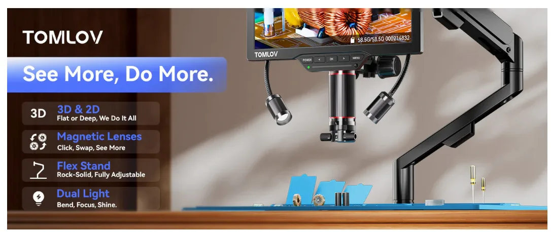
These microscopes combine advanced optics with powerful software to generate detailed 3D models or depth maps on a computer screen. The “3D” is often reconstructed from 2D image data.
- How They Work: A common method involves focus variation or z-stacking. The microscope captures a series of 2D images at incrementally different focal planes as it moves vertically through the sample. Software then processes these images, identifying the in-focus points at each depth, and reconstructs a high-resolution 3D model or a depth-encoded image.
- Ideal Applications:
- Industrial Metrology & Quality Assurance: Precisely measure surface roughness, feature dimensions, and defect volumes.
- Failure Analysis: Analyze fracture surfaces, corrosion pits, or wear patterns in great detail.
- Research & Development: Characterize new materials, coatings, or micro-components.
- Documentation: Create highly detailed, fully focused 3D images for reports and presentations.
- Key Consideration: The “3D” is viewed on a screen and is a digital reconstruction, not direct optical depth perception like a stereo microscope. However, it offers superior measurement capabilities and resolution for surface topography.
Confocal Microscopes: Unveiling Internal Structures
Confocal microscopy is a sophisticated technique primarily used in life sciences and materials science to obtain high-resolution 3D images of samples, particularly thick or scattering specimens.
- How They Work: Confocal microscopes use a laser to scan a point across the specimen. Crucially, they employ a pinhole aperture in front of the detector to block out-of-focus light from above and below the focal plane. This creates an “optical section” – a very thin, sharp 2D image. By collecting many such optical sections at different depths (a “z-stack”), software can reconstruct a stunning 3D image of the specimen’s internal structure.
- Ideal Applications:
- Cell Biology & Neuroscience: Imaging fluorescently labeled cells, organelles, and neural networks in 3D.
- Developmental Biology: Studying tissue development and growth in whole organisms.
- Materials Science: Analyzing the internal structure of polymers, composites, or ceramics.
- Live Imaging: Observing dynamic processes within living cells in a 3D context.
- Key Consideration: Confocal microscopes are significantly more complex and expensive than stereo or basic digital microscopes, requiring specialized training and sample preparation.
Beyond the Common: Holographic & Electron Microscopes
While less common for general inquiries, these represent the cutting edge of 3D imaging at the micro and nano scale.
- Holographic & Light-Field Microscopes: These innovative systems capture more comprehensive light information from the sample, often in a single shot, allowing for computational reconstruction of the 3D volume. They are pushing boundaries in high-speed 3D imaging of dynamic biological processes.
- 3D Electron Microscopes: For visualizing structures at the nanoscale (atoms and molecules), electron microscopes are essential. Techniques like electron tomography involve tilting a sample and taking a series of 2D images, which are then computationally combined to create a 3D reconstruction of the sample’s electron density. This is akin to a CT scan, but at an incredibly fine resolution.
- Key Applications: Nanomaterials research, advanced biological ultrastructure analysis, and semiconductor inspection.
By answering these questions, you’ll be well on your way to understanding which type of 3D microscope truly fits your needs!
Frequently Asked Questions (FAQs)
Q1: Do 3D microscopes require special glasses to see the 3D effect?
A1: Only some very specific, often older or specialized, 3D display systems might require glasses. Most commonly, stereo microscopes provide natural 3D vision through two eyepieces, while 3D digital microscopes display a rotatable 3D model on a standard monitor, which doesn’t require glasses.
Q2: Can a regular 2D microscope be converted into a 3D microscope?
A2: A traditional 2D optical microscope cannot become a true stereo microscope for direct 3D viewing. However, some digital camera attachments for 2D microscopes can use software techniques like “z-stacking” to create a 3D reconstruction or extended depth of field image from multiple 2D captures, giving a 3D-like effect on a computer screen.
Q3: What’s the biggest advantage of a 3D microscope over a traditional 2D one?
A3: The biggest advantage is the ability to perceive or measure height and depth. This is crucial for tasks requiring hand-eye coordination (like soldering), for accurate surface metrology (like measuring scratch depth), or for understanding the complex internal architecture of biological samples.
Q4: Are 3D microscopes expensive?
A4: The cost varies dramatically depending on the type and capabilities. Basic stereo microscopes for hobbyists can cost a few hundred dollars. High-end 3D digital microscopes or advanced confocal systems for research can range from tens of thousands to hundreds of thousands of dollars.
Q5: Can I get 3D measurements with all types of 3D microscopes?
A5: While all 3D microscopes provide some form of depth information, the precision and type of measurement vary. 3D digital microscopes excel at quantitative surface metrology (roughness, height, volume). Confocal and electron microscopes provide highly precise measurements of internal structures based on their volumetric data. Stereo microscopes offer qualitative depth perception, but their measurement capabilities are more basic, typically for linear dimensions.


